Mohd Farid

Mohd Farid
Showing posts with label ECG Interpretation. Show all posts

Saturday, March 14, 2020

You Ask I Find The Answer-ECG Interpretation: Infarction-Injury Part IIIb ST Depression

Infarction

Myocardial Infarction Triad

Injury
(means acute or recent)

ST Depression


The ST segment may become depressed under certain circumstances or conditions. 

During an angina attack, the ST segment may be temporarily depressed.

A subendocardial infarction, an infarct that does not extend through the full thickness of the left ventricular wall, will depress the ST segment.

Positive Stress Test


When a patient with narrowed coronaries exercises, the myocardium demands more blood flow than its arteries can deliver. A stress or exercise test will record depression of the ST segment on EKG when such a patient is exercised.

Digitalis

Digitalis can cause depression of the ST segment, however it has a unique, unforgettable appearance. 


Digitalis causes a gradual downward curve of the ST segment, to give it the appearance of Salvador Dali's mustache. Notice that the lowest portion of the ST segment is depressed below the baseline.

Digitalis produces a unique, gradual downward curve of the ST segment; this is the classical "digitalis effect". 

To identify the classical pattern of digitalis effect, you should observe a lead with no demonstrable S wave. The downward portion of the R wave gradually thickens as it curves down into the ST segment, which is usually depressed. The downward limb of the R wave has a gentle, curving slope that gradually blends into the depressed ST segment. Look for it the next time you have a patient on a digitalis preparation. 

Subendocardial Infarction


Subendocardial infarction causes flat depression of the ST segment; however, any significant ST depression (in leads where the QRS is upright) indicates compromised coronary blood flow until proven otherwise.

Subendocardial infarction (often referred to as subendocardial injury) is identified by flat ST segment depression, which may be either horizontal or down sloping.

Subendocardial infarction, a type of "non Q wave infarction" involves only a small area of myocardium just beneath the endocardial lining.  Classical myocardial infarction is said to be transmural; that is, the full thickness of the left ventricular wall is damaged in the infarcted area. Even though subendocardial infarction involves only a small area of the myocardium, it must be respected as a true MI that requires appropriate care. A subendocardial MI may enlarge or extend and become more life threatening.

Any patient with acute ST depression or elevation, particularly if it persists, should have an immediate, complete workup including cardiac enzymes. 

Credit: Dale Dubin-RapidInterpreatation Of EKG 

You Ask I Find The Answer-ECG Interpretation: Infarction-Injury Part IIIa

Infarction

Myocardial Infarction Triad

Injury
(means acute or recent)


Injury indicates the acuteness of an infarct. Elevation of the ST segment denotes "injury" sometimes called the "current of injury".

The ST segment is that section of baseline between the QRS complex and the T wave. The ST segment contains.

Elevation of the ST segment signifies "injury". The ST segment may be elevated only slightly, or as much as one or more milimeters above the baseline.

ST segment elevation tells us that a myocardial infarction is acute. It is the earliest consistent sign of infarction to record on EKG.

Angina with exertion, "Prinxmetal's angina, can cause transient ST elevation in the absence of an infarction.

ST Elevation

If there is ST elevation, this indicates that the infarction is acute. ST elevation, alone, can indicate an infarction.

Once you have made a diagnosis of infarction, it is important to know whether the infarction just occurred and needs immediate treatment, or if the infarction is old, maybe years old.

The ST segment rises above the baseline with an acute infarction, in fact it is usually the earliest EKG sign of an infarction. With time, the ST segment returns to the baseline. 

If the ST segment is elevated without associated Q waves, this may represent non-Q wave infarction, which is usually a small infarction that may herald an impending larger infarct. Significant ST changes require enzyme studies and close scrutiny.

A ventricular aneurysm (the outward ballooning of the wall of ventricle) can cause persistent ST elevation in most of the chest leads; but in this case, the ST segment does not return to the baseline with time. Pericarditis (next page) produces a unique type of ST segment elevation that may also elevate the T wave off the baseline.

Brugada Syndrome-ST Elevation


RBBB pattern QRS with ST elevation in V1-V3

Can cause sudden cardiac arrest (in absence of coronary obstruction)

Brugada syndrome is a hereditary condition that can cause sudden death in individuals without heart disease. It is characterized by Right Bundle Branch Block with ST elevation in leads V1 to V3. Look for it; this malady is not rare.

Sudden cardiac death (cardiac arrest ) can occur spontaneously in patients with Brugada syndrome.

In Brugada syndrome there is RBBB and ST elevation in leads V1 to V3. The elevated ST segments have a peculiar, peaked downsloping shape, particularly in V1 and V2.

Brugada syndrome is a familial condition caused by dysfunctional cardiac Na+ (sodium) channels. Prophylaxis against the deadly arrhythmias requires ICD implantation in order to immediately treat cardiac arrest (usually ventricular fibrillation). 

This syndrome is responsible for nearly one half of the sudden deaths in healthy young individuals without structural heart disease.

Pericarditis-ST Elevation


With pericarditis, the ST segment is elevated and usually flat or concave. The entire T wave may be elevated off the baseline. 

Pericarditis is inflammation of the membrane (pericardium) that surrounds the heart. Pericarditis may be caused by a virus, bacteria, cancer, or other sources of inflammation, including myocardial infarction.

Pericarditis can elevate the ST segment. It usually produces an elevated ST segment that is flat or slightly concave (middle sags downward). This resolves with time.

Pericarditis seems to elevate the entire T wave off the baseline; that is, the baseline gradually angles back down (often including the P wave) all the way to next QRS.


The characteristics shown in the left illustration are found in lead in which the QRS is usually mainly negative (like the right chest leads). The pattern shown in the right illustration is seen in leads where the QRS is mainly positive (such as the lateral and inferior limb leads). Sometimes PVC's are produced.

You Ask I Find The Answer-ECG Interpretation: Infarction-Ischemia Part II

Infarction

Myocardial Infarction Triad

Ischemia


Ischemia (decreased blood supply) is characterized by inverted T waves.

Ischemia means reduced blood supply (from the coronary arteries); the ischemic area is at the periphery of the infarct.

The characteristic sign of ischemia is the inverted T wave. It may vary from a slightly inverted to a deeply inverted T wave.

Inverted T waves may indicate ischemia in the absence of myocardial infarction. Coronary blood flow can decrease without producing an infarction.

Cardiac ischemia alone can cause chest pain known as angina, which is usually associated with transient T wave inversion.


The typical ischemia T wave is symmetrically inverted.

You should check every EKG that you read for T wave inversion. Since the chest leads are nearest the ventricles, T wave changes are most pronounced in these leads. Always run down V1 to V6 (as well as the limb leads) and check for T wave inversion to see if there is diminished coronary flow.

The T wave of ischemia is both inverted and symmetrical; that is, the right and left sides of the inverted T wave are mirror images.

In adults flat (nonexistent) T waves or minimal T wave inversion may be a normal variant in any of the limb leads (frontal plane). However, any T wave inversion in leads V2 through V6 is considered pathological. Marked T wave inversion in leads V2 and V3 the hallmark of Wellens Syndrome, alerts us to stenosis of the anterior descending coronary. 

Credit: Dale Dubin-RapidInterpreatation Of EKG

Friday, March 13, 2020

You Ask I Find The Answer-ECG Interpretation: Infarction Part I

Infarction

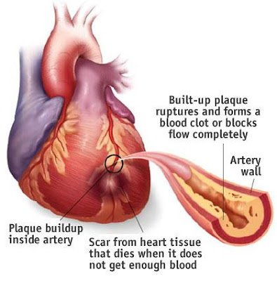
Myocardial Infarction results from the complete occlusion of a coronary artery. The infarcted area of myocardium becomes necrotic (dead), so it cant depolarize or contract. 


Although the heart's chambers are filled with blood, the myocardium's own blood supply is provided exclusively by the coronary arteries. A coronary artery can be gradually narrowed by lipid deposits that become atheromatous plaque beneath the intima lining of the vessel. The intima may eventually rupture, exposing the plaque to the blood within the artery. This initiates the immediate formation of a clot (thrombus). The vessel, already narrowed by the plaque, becomes totally occluded by the thrombus. Instantly the infarcted area of the ventricle (without a blood supply) becomes necrotic. Ventricular foci in the hypoxic area around the infarct become very irritable, this can produce deadly ventricular arrhythmias. 

Myocardial Infarction implies the complete occlussion of a coronary artery, which we can diagnose with the EKG. The electrocardiogram will also tell us which coronary artery or coronary branch is occluded, and it can even reveal any blocks in the ventricular conduction caused by the infarction. By careful interpretation of the EKG, we can also determine if a coronary vessel is narrowed, rendering a decreased blood supply to the heart. Practical livesaving knowledge. 


Myocardial infarction is due to the occlussion of a coronary artery supplying the left ventricle, so an area of the heart is without a blood supply and suffers necrosis.

The terms "myocardial infarction", "coronary occlusion" and "heart attack" refer to the same serious phenomenon. 

The heart derives its own blood supply from the coronary arteries, so when a coronary artery or one of its major branches is occluded, an area of the myocardium is without blood supply. 

The infarcted (necrotic) area is primarily in the left ventricle; deadly arrhythmias may result.

We understand that the coronary arteries also supply the right ventricle, so there is often some involvement of the right ventricle,. But since most of the critical problems originate in left ventricular infarcts, myocardial infarction is usually conceptualized in term of the left ventricle. 


Commonly, the thick left ventricle that suffers myocardial infarction

The left ventricle is the thickest chamber of the heart; so if the coronary arteries are narrowed, the left ventricle (which uses the greatest blood supply) is the first to suffer from an obstructed coronary artery.

Blood is pumped to all parts of the body by the powerful, thick, left ventricle.

When we describe infarcts by location, we are speaking of an area within the left ventricle. Coronary arteries to the left ventricle usually send smaller branches  to other regions of the heart, so an infarction of the left ventricle can include a small portion of another chamber.


The necrotic infarcted area of the ventricle (that has no blood supply) is electrically dead and cannot depolarize.

Infarctions usually involve an area of the wall of the left ventricle.

An area of infarction cannot be depolarized because the cells there are without s blood supply, so they are necrotic (functionally dead).

This necrotic infarcted area produces an electrical void, while the rest of the heart (with an adequate blood supply) functions as usual. The infarcted region does not depolarize, so it does not contract, thereby impairing the muscular function of the left ventricle. Also, hypoxic ventricular foci nearby are often the source of serious ventricular arrhythmias. 


The myocardial infarction triad is "ischemia", "injury" and "necrosis", but any of the three may occur alone.

Necrosis (death) of a ventricular region produces dead mayocardial cells that cannot depolarize.

The myocardial infarction triad is the basis for recognizing and diagnosing a myocardial infarction.

The word hypoxia means decreased oxygen; in the heart, it is usually caused by ischemia, which literally means reduced blood supply (diminished blood flow).

"Ischemia", "injury" and "necrosis" need not all be present at once in order to establish the diagnosis of myocardial infarction. Routine EKG interpretation requires checking these infarction criteria.

Credit: Dale Dubin-RapidInterpreatation Of EKG