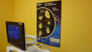
Imaging For
(Speckle) Strain With Intent To Analyze In Room
-Don’t change data flow, leave in internal hard
drive/dicom server.
-Turn off dual focus – move focus from bottom of screen to
middle of screen.
-Check depth and
width for each of the three apical windows (Apical Long Axis, Apical 4 Ch,
Apical 2 Ch) for LV before start acquiring.
-All three views from each view have to have identical
depth/width/frame rate.
-Frame rate has to be >40 fps, if not turn frame rate
knob one click clockwise.
-Do the best you can in the room to acquire clips with
very similar heart rates.
-Watch to see if rate changes with held breath in or out.
-If Atrial
Fibrillation/Irregular HR, increase clip storage from 3 beats to 5 beats or
more.
-If irregular HR,
look for similar R-R interval for each apical view and isolate cardiac cycle
with number of cycles knb. Change to one cycle, then with cycle select knob pick
which cardiac cycle you want work with before assigning view label (Apical Long
Axis, Apical 4 Ch, Apical 2 Ch).
-Analyze Apical
Long Axis first. If not tracking correctly, push the “Recale” butto. Next view
is about do we want to analyze only to aortic valve closure or later. Don’t
change a thing, just click on kidney button.
-Before moving on
to next view, click on quad view button. Make sure mark on waveforms is where
it should be. If mark is on the early systolic upward part of waveform, move to
downward part of waveform in late systole by clicking on mark. The waveform
turns a bold color, then move to desired location (machine will not allow to move
past valve valve closure), also in this view:note numbered results on 2D
image-clip image.
-Do same for
Apical 4 Ch then Apical 2 Ch view.
-May see message
about aortic valve closure problems. If so, you will not get a bull’s eye
composite view, but with 3 clipped quad views you will have all strain numbers
except the apical cap.
Speckle Strain Is
Done
-Measure LVOT for
event timing.
-Acquire narrowed
sector view of inferoseptum with TDI on, then analyze Doppler based strain of
basal segment.
-No need to acquire
the three full TDI views for backup; speckle strain is already done
-Continue on with
the normal echo exam
If Images Are
Difficult Or Having Wall Tracking Problems
-End your exam by
clicking on New Patient; change data flow to interal hard drive.
-Pick study you
were working on, click on select, not create, continue-No.
-Acquire strain
study to be sent out to GE echo pac. All images, including the three apical
views, full TDI and LVOT Doppler.
-Send out to echo
pac, change data flow back to internal hard drive dicom/server.
-Pick study you
were working on, click on select, not create, continue-No.
-Finish regular
echo examination.
Heart Rate And
Strain Waveform Issue On GE Echo Pac
-If irregular HR,
look for similar R-R interval for each apical view, isolate with number of
cycles arrows, and change to one cardiac cycle. Then with cycle select arrows,
pick which cardiac cycle you want to work with before assigning label
(ALAX,4Ch, 2 Ch).
-Still having HR
problems?
-Click on quad
view (after successfully analyzing each individual view) to get the strain
info.
.
-You will see the
strain results for each wall from that view.
-Do the same for
each view.
-You will not get
a composite bull’s eye view.
-You will not get
strain of the apical cap.
Strain (Speckle)
Final Check
-Check to see if
strain waveforms make sense in the 3 strain waveform and bull’s eye view.
-Make sure mark on
waveform is where it should be. If mark is on the early systolic upward part of
waveform, move to most downward part of waveform in late systole by clicking on
mark. The waveform will turn a bold color, then move to desired location
(machine will not allow to move past valve closure).
-For cases where
more than 2 segments cannot be visualized, it also cannot be evaluated by 2D
speckle strain, Doppler strain should be used.
Strain
Updates
-Make sure QRS complex is highest upward deflection
not P or T waves.
-Measure strain only until aortic valve closure for
Doppler and speckle are determined.
-Wall segments that are not tracking by speckle
strain need to be analyzed by Doppler stra
-Frame rate needs to be above 200 fps for Doppler
based strain, narrow sector.
-When analyzing Doppler strain make sure strain start
bracket is at start of QRS complex, not start of P wave.
-General strain news : with
the Vivid E9 Echo Machine we cannot manipulate the waveforms until all three
views are completed and you are at the bullseye with three quad views. With
Doppler based strain on Vivid E9 click on the strain button before you click on
the Q analysis button. Enter strain numbers into your system if necessary from
bull’s eye not from the stored quad views, the results will be different if
entered from quad view rather from Bullseye. After analyzing the 2 Chamber View
remember to store the 2 Chamber Quad View, the Vivid E9 bypasses this step.
LV
Strain Acquisition And Analysis
-Turn off dual focus-move focus from bottom of screen
to middle of screen.
-Check depth and width for each of the three apical
windows for LV before you start acquiring-all three views from each view have
to have identical depth/width/frame rate.
-Frame rate has to be > 40 fps-if not turn frame rate
knob one click clockwise.
-Do the best you can in the room to acquire clips
with very similar heart rates.
-Watch to see if rates changes with held breath in or
out.
-If Atrial Fibrillation or Irregular Rhythm, increase
clip from 3 beats to 5 beats or more.
-If irregular HR, look for similar R-R interval for
each apical views and isolate cardiac cycle with number of cycles knob. Change
to one cycle, then with cycle select knob pick which cardiac cycle you want
with before assigning view label (Apical Long Axis, Apical 4 Ch, Apical 2 Ch).
-Analyze Apical Long Axis View first. If not tracking
correctly, push the Recalc button (up by screen). Next view is about do we want
to analyze only to aortic valve closure or later. Don’t change a thing, just
click on kidney button.
-Before moving on to next view, click on quad view
button. Make sure mark on waveform is where it should be. If mark is on the
early systolic upward part of waveform-move to downward part of waveform in
late systole by clicking on mark. The waveform turns a bold color, then move a
desired location (machine will not allow to move past valve closure).
-Also in this view: note numbered results on 2D
image-clip, save quad view.
-Do same for Apical 4 Ch and Apical 2 Ch Views.
-Measure LVOT for event timing, Aortic valve opening
and Aortic valve opening.
-Acquire narrowed sector view of inferoseptum with
TDI on, then analyze Doppler based strain of basal segment and save.
RV Strain
Goal
-Quantification
of right sided longitudinal contractility in pulmonary hypertension patients.
-Patients
undergoing an initial or subsequent PHTN echo will have longitudinal systolic
strain recorded from the free wall of the right ventricle.
-Unlike the
LV, the predominant orientation of muscle fibers in the RV is in the
longitudinal plane.
-Measures of
transverse/radial function do not directly reflect the major component of RV
systolic function.
RV Strain Updates
With the new
Vivid E9 just bring the RV center, analyze by calling it the Apical Long Axis
View. That should be the only view needed. Pay close attention, the waveforms
for the RV free wall will now be different than they would be when we analyzed
by calling the RV the 4 Ch view.
General
strain news: with the Vivid E9 we cannot manipulate the waveforms until all
three views are completed and you are at the bullseye with three quad views.
With Doppler based strain click on the strain button before you click on the Q
analysis button.
RV Strain Image Acquisition
We don’t
need Apical Long Axis View with the Vivid 7, machine will estimate aortic valve
closure (sensing change in volumes), go directly to 4 Ch view
-Turn off dual focus-move single focus to
approximately mid screen or to where image is most optimized.
-Narrow image sector and use tilt function (if
needed) to isolate the RV.
-Frame rate has to be > 40 fps.
-Best results may come from a more lateral apical
window
.
-Walls don’t need to be parallel to ultrasound beam
due to nature of speckle analysis.
-Make sure depth is set appropriately so as not to
cut off the lateral annulus.
-Insert point at base septum, base lateral wall, and
apex.
-Place base septum and base lateral wall points at
annulus.
-Pull points down to annulus, not beyond annulus.
-Place septal point at tricuspid insert side, not
mitral insert side.
-Make sure apical point is at true apex.
-Should track the blood tissue interface, may need to
move/increase ROI width to track.
-Will try our best to analyze with speckle strain.
-Problem wall will most likely be basal lateral wall.
-Septum does not need to track, not reporting septum.
-If unsuccessful, will acquire with Doppler strain.
To Analyze The RV Free Wall Doppler
Based Strain
·
Before Doppler strain, do event timing using RVOT,
instead of LVOT.
·
Measurements→Event Timing→Ao Valve Opening-Ao Valve
Closing
·
Wall does need to be parallel with ultrasound beam
·
Frame rate needs to be > 200 fps
RV Strain Data
-RV Basal Lateral Peak %
-RV Mid Lateral Peak %
-RV Apical Lateral Peak %
-RV Lateral wall averaged peak systolic strain %
-Best to go to graphics pull down menu to strain
(longitudinal systolic), as measurement page may be going away.


No comments:
Post a Comment