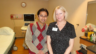
Echo Technologist
Echo
Technologist/Sonographer will be doing echocardiogram only. They don’t do
anything else than echocardiogram. Echo technologist will performs cardiac ultrasound procedures such as M‑mode,
2‑D, Doppler and Color Flow Doppler utilizing a variety of equipment and
techniques and makes the necessary measurements required for the interpretation
to obtain accurate, high quality results (for example, muscle wall thickness,
diameter of chambers, volume of chambers, valve areas, blood flow velocity and
direction).
This
centre have specific technologist for each area including stress test, nuclear
medicine, electrophysiology study, cath procedure and others. Sonographer will
be at cath lab during specific procedure only including TEE, TAVI, Mitral Clip,
TASH, Emergency Pericardiocentesis and other related echo procedure. In Stress
Echo procedure there will be stress technologist that do the stress test and
help in injecting contrast agent at peak exercise so Echo Technologist will be
focusing on grabbing good echo images.
Transthoracic
Echocardiography
For
Transthoracic Echocardiography, Echo Technologist will get all patient
demographic data (height and weight) , patient medical record and check for
blood pressure before they start the echo test. This data may be valuable to
get general idea about patient echo diagnosis. Each Echo test will be 75
minutes slot. So they will be enough time to do a complete echo study that may
include Biplane Simpson, 3D Echocardiography, Strain Imaging, Strain Rate, and
Dyssynchrony Study if necessary. In
Aortic Stenosis case, they required to use Pedof probe to get good Aortic Valve
Doppler tracing in evaluation of Aortic Stenosis. It is part of ICAEL requirements.
Cardiac Specialty Centre
St.
Luke’s also recently opened Cardiac Specialty Centers that offer the latest
treatments and research for hypertrophic cardiomyopathy, adult congenital heart
disease, Marfan’s and aortic disorders, and valvular heart disease. A.
Jamil Tajik, MD, an internationally recognized expert, heads the centers
and leads a multidisciplinary team of specialists who coordinate patient care.
These complex conditions can be fatal if undiagnosed.
Aurora
Health Care wants to be a leader in the treatment of complex heart conditions.
Their specialty heart centers serve patients with hypertrophic cardiomyopathy,
adult congenital
heart disease, Marfan’s
syndrome and valvular heart disease.
They offer the highest level of care in the hands of experts utilizing advanced
diagnostic and therapeutic technologies. In fact, their specialty services for
adult congenital heart disease and hypertrophic cardiomyopathy are the only
dedicated specialty centers in the state.
An
accredited echocardiography laboratory requires the interpreting physicians and
practicing sonographers to be adequately trained and experienced to interpret
and perform echocardiograms. So they will Consultant or Cardiologist that will
be read, review and finalized echo report in their respective review room. At
Cardiac Specialty Centre, Cardiologist can review ongoing echo procedure at
their review room without any waiting for transferred data to prosolv.
Cardiologist can interfere and request any additional information during that
echo test.
Ejection Fraction
Ejection
fraction is a measurement of the percentage of blood leaving your heart each
time it contracts. During each heartbeat cycle, the heart contracts and
relaxes. When your heart contracts, it ejects blood from the two pumping
chambers (ventricles). When your heart relaxes, the ventricles refill with
blood. No matter how forceful the contraction, it doesn't empty all of the
blood out of a ventricle. The term "ejection fraction" refers to the
percentage of blood that's pumped out of a filled ventricle with each
heartbeat. During an echocardiogram, sound waves are used to produce images of
your heart and the blood pumping through your heart.
Aurora
did not use Teich Method (M-Mode) anymore to measure Ejection Fraction. They
made the 2D Biplane EF quantification as a compulsory method to calculated EF.
Now, if they all unable or have inability to visualize at least 2 myocardial
segments of the left ventricle, they will use contrast agent to make 2D Biplane
EF quantification. They are planning to make 3D EF Measurement as a compulsory
method next year. The echo technologist is now practicing to make the 3D EF
quantification more reproducible and reliable to do when the time come.
Contrast Agent
The cardiac
sonographer has the responsibility to carry out the physician’s order for the
performance of the examination and therefore initially determines the overall
technical quality of the echocardiogram. Thus this person is often in the
primary position to identify the need for contrast enhancement. It is well
established that in up to 20% of resting studies, the endocardial border
definition of the left ventricle is suboptimal. This has been defined as the
inability to visualize at least 2 myocardial segments of the left ventricle.
Suboptimal visualization of certain segments, such as the anterior or lateral
walls, may be even worse during stress echocardiography. The enhancement of
left ventricular endocardial border definition can be facilitated by using
transpulmonary contrast agents. This centre widely used Contrast Agent
(Definity) to help to gain good image and diagnosis for the patient that with
poor echo window and give echo test more reproducible value in each echo test
that they have done. It will be helpful in test such as stress echocardiogram
because we can see more better endocardial representation with contrast agent.
Transesophangeal
Echocardiography
There
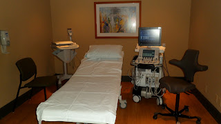
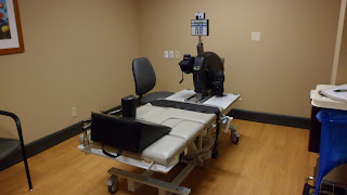
is respective Echo Room for Transesophangeal Echocardiography, Transthoracic
Echocardiogram and Stress Echocardiogram. Transesophangeal Echocardiography
procedure will be done by cardiologist assisted by Echo Technologist and
accompanied by Registered Nurse (RN) in case needed used of medication
especially for sedate patient procedure begin. Registered Nurse will monitored
patient vital sign (SPO2, ECG and Blood Pressure). Sonographer will handle
knobology and other specific modalities depends on protocol and cardiologist
interest.
This
centre also involve in Transcatether Valve intervention. They have being doing
very well in Mitral Clip Procedure and Transcatether Aortic Valve Implantation
(TAVI). Transesophangeal Echocardiography is useful in this procedure that will
be done in Hybrid Operation Theater. Echo Technologist also will be doing
Transesophangeal Echocardiography at EP Cath Lab for Cardioversion procedure
and Transeptal Puncture for RFA. Echo Technologist will be accompanying by Echo
Cardiologist.
Transcatheter Aortic
Valve Implantation
Transcatheter aortic valve
implantation (replacement) is an investigational treatment for severe aortic
stenosis. The purpose of the clinical trial is to learn whether transcatheter
aortic valve implantation is a safe and effective treatment option for certain
patients with severe aortic stenosis. With transcatheter aortic valve
implantation, an artificial aortic heart valve attached to a wire frame is
guided by catheter to the heart. Once in the proper position in the heart, the
wire frame expands, allowing the new aortic valve to open and begin to pump
blood.
In This procedure, echo technologist
will assist Echo Cardiologist in performing TEE on the patient before
procedure, during procedure and post procedure. Before procedure, echo team
will assess aortic valve pathology, baseline Aortic Valve Mean Gradient, Aortic
Valve Peak Gradient, Aortic Valve Peak Velocity, Severity Of Aortic Regurgitation,
and Severity Of Mitral Regurgitation. During procedure, Echo team will be
assessing the position of implantable AV if required by interventional
cardiologist. After procedure, echo team will assess aortic valve, Aortic Valve
Mean Gradient, Aortic Valve Peak Gradient, Aortic Valve Peak Velocity, Severity
Of Aortic Regurgitation (Paravalvular Or Transvalvular), any evidence of
effusion (Pericardial or Pleural) and any evidence of LVOT Obstructions
(Systolic Anterior Motion).
Sonographer Credentialing
And ICAEL
Almost
all sonographer at Aurora St Lukes Medical Centre are registered under specific
organization. American Registered Diagnostic Medical Sonography (ARDMS) will
give title Registered Diagnostic Cardiac Sonographer (RDCS) , Cardiovascular
Credentialing International (CCI) will give title Registered Cardiac
Sonographer (RCS) and American Society Of Echocardiography (ASE) will give
title Fellow Of American Society Of Echocardiography (FASE).
This will required
assestment and examination that must be pass by all respective candidates. Aurora
St Lukes Medical Centre echocardiography laboratory also is accredited by The
Intersocietal Commission For The Accreditation Of Echocardiography Laboratories
(ICAEL) that can improved services. An accredited echocardiography laboratory
requires the interpreting physicians and practicing sonographers to be
adequately trained and experienced to interpret and perform echocardiograms. So
they will Consultant or Cardiologist that will be read, review and finalized
echo report in their respective review room.
Online Reporting
For
echocardiography reporting, the echo technologist will do the online reporting
on Prosolv System and charge every patient by himself. Echo technologist will
do all the measurement in Prosolv for cardiologist reference. The conclusion
for the echocardiography assessment will be done by the Cardiologist himself.
All decision making will depends on the echocardiologist to decide.


No comments:
Post a Comment